


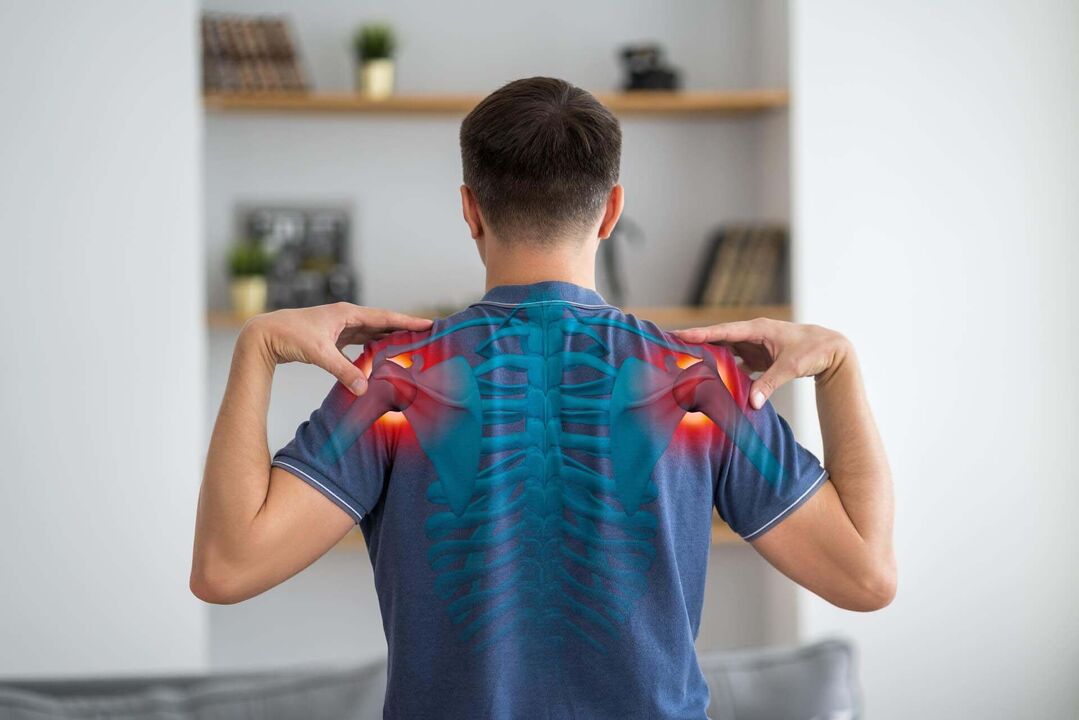
Arthrosis of the shoulder joint is a dystrophic change of the cartilaginous plate covering the articular surfaces of the joint, with subsequent involvement of the underlying bone.
About the disease
This disease affects not only the cartilage layer and the bone under the cartilage. The pathological process gradually affects the joint capsule and the ligament apparatus, the synovium, the musculotendinous compartment, and the subacromial region.
At a certain stage, arthrosis of the shoulder joint can lead to the development of osteoarthritis. This condition is characterized by the following symptoms: chronic pain, reduced range of motion in the joint, intra-articular cracking during rotation. People over the age of 40 are most often exposed to this transformation.
The main symptoms of arthrosis of the shoulder joint are pain and limited mobility of the arm. To confirm the diagnosis, imaging methods are informative - ultrasound and X-ray scanning, computed tomography and magnetic resonance imaging.
According to clinical recommendations, the initial stage of the disease is treated with conservative methods, and in the later stages, when there is significant damage to the cartilage layer and the patient's self-care is impaired, joint replacement is required.
Types of arthrosis of the shoulder joint
According to the classification, the following types of arthrosis of the shoulder joint are distinguished:
- primary arthrosis, in the development of which genetics plays a major role, and even the most thorough examination does not allow identifying the most significant cause of the disease;
- secondary arthrosis, which is the result of adverse factors affecting the joint (trauma, endocrine diseases, damage to joint anatomy).
Doctors judge the speed of progression of the pathological process based on the extent of the disease. The more aggressive the process, the faster the destruction of the articular cartilage and the involvement of the underlying bone. From a morphological point of view, arthrosis of the shoulder joint has 6 degrees:
- first degree - the cartilage matrix swells and disintegrates, but the integrity of the surface zone of the cartilage is not yet damaged;
- second degree – the cells of the cartilage tissue located in the deep layers are affected, the surface plate of the cartilage is damaged;
- third degree - vertical cracks appear on the cartilage plate;
- fourth grade - the surface zone of the cartilaginous plate gradually peels off, erosion defects are formed, and cystic cavities appear in the underlying bone;
- fifth grade - at this stage, the underlying bone is visible;
- sixth degree - the subchondral zone thickens significantly, the cysts become more pronounced, and marginal bone growth appears.
Symptoms of arthrosis of the shoulder joint
The main clinical signs of shoulder arthrosis are pain, stiffness of the joint up to complete loss of mobility, and deformation of the joint.
Distinctive features of deforming arthrosis pain are:
- appearance at the beginning of flexion, extension or rotation;
- increased during physical activity;
- nocturnal character due to stagnation of venous blood in the intraosseous channels;
- the presence of blockages - a sudden blockage in the joint due to the separation of separated osteochondral fragments, which are placed between the joint surfaces;
- weather dependence - the pain increases with changes in the weather (in wet and cold climates, the pain becomes more intense).
Arthrosis is a chronic pathology. In the initial stage of the disease, the pain appears intermittently (during the exacerbation of the disease). The speed of the progression of the pathology is determined by the timeliness of the start of the treatment and the appropriateness of the lifestyle modification. Shoulder pain becomes chronic if it lasts for 6 months or more. Acute pain becoming chronic indicates the progress of the pathological process.
Causes of arthrosis of the shoulder joint
The causes of arthrosis of the shoulder joint can be divided into 2 groups:
- modifiable - correction is possible;
- cannot be modified - their action cannot be influenced.
Non-modifiable factors that can increase the risk of developing arthrosis of the shoulder joint include:
- gender - up to the age of 50, women are less susceptible to the disease than men; after about 50 years, the prevalence of pathology among representatives of both sexes will be approximately the same;
- the age of the person - the older the patient, the greater the risk (and from the age of about 30, the degeneration process in the cartilage tissue takes place faster than the regeneration process, which creates the prerequisites for the development of the disease);
- congenital disorders of the structure of the shoulder - excessively increased mobility (hypermobility), connective tissue dysplasia (usually the articular cartilage is represented by type 2 collagen fibers, replaced by dysplasia, a less durable type of collagen), instability of the articulation;
- genetic characteristics - hereditary predominance of type 2 collagen, interleukin-1 and interleukin-2 gene polymorphism.
Modifiable risk factors for deforming arthrosis of the right or left shoulder joint:
- traumatic joint damage;
- excessive physical activity (strength sports and martial arts, including weight bench pressing);
- obesity - in the case of shoulder joint arthrosis, it is not the increase in mechanical load that is important, but the metabolic changes occurring in the connective tissue, e. g. the chronic inflammatory condition accompanying obesity;
- weakness of the muscle ligament of the shoulder joint, especially in people who perform precise activities with their hands (jewelers, dentists, secretaries, writers);
- lack of vitamin D, which is actively involved in maintaining the health of the musculoskeletal system;
- a diet low in vitamin C, which is an important link in the body's calcium-phosphorus metabolism;
- hormonal imbalance - thyroid disease, diabetes, etc. ;
- smoking – both active and passive.
In shoulder joint arthrosis, the main target of the pathological process is the articular cartilage, subchondral bone and synovium. In the affected cartilage, the synthesis of proteoglycans decreases, fragmentation and cracking of the disc is observed, which exposes the underlying bone. An increase in the non-physiological load of the bone leads to its compaction, the appearance of cysts and osteophytes (marginal growths).
Diagnostics
The examination of a patient with pain in the shoulder joint should begin with X-rays. To examine the joint in detail, it is important to scan in several projections. Pictures can be taken in direct projection, internal and external rotation. To assess the soft tissue formations of the joint, especially in the early stages of arthrosis, the ultrasound examination of the joint is the most informative. If the diagnosis remains unclear, magnetic resonance imaging/computed tomography of the joint is recommended. In the next stage, the preservation of the functions of the articulation is evaluated.
Expert opinion
All morphological formations of the joint are involved in the pathological process. The main symptom of osteoarthritis is joint pain, which is caused not only by arthritis, but also by bone damage (osteitis, periostitis), involvement of the periarticular soft tissues (tendinitis, tenosynovitis, myalgia, enthesopathies, stretching of the joint capsule). , degeneration of the menisci and involvement of the neurosensory system (for example, irritation of nerve trunks by large osteophytes). Therefore, the treatment starts as soon as possible, e. g. lifestyle modification, the more effective the control of the occurrence of pain will be.
Treatment
At the initial stage of the pathological process, the treatment of arthrosis of the shoulder joint is carried out with conservative methods, and with severe degeneration of the articular cartilage, surgical intervention (endoprosthesis) is indicated.
Conservative treatment
In the period of exacerbation of the process, the first priority is pain relief. Non-steroidal anti-inflammatory drugs are most often used for pain relief. They can be applied locally (in the form of creams and ointments), injected into the joint cavity, or used systemically (tablets, intramuscular injections). In some patients, the pain may be so severe that a short course of corticosteroids may be used to relieve the pain.
Intra-articular injection of hyaluronic acid or plasma, including those enriched with platelets, can stimulate the cartilage plate and promote its renewal (this treatment is considered pathogenetic). These injections help speed up the synthesis of collagen and elastin fibers, which form the basis of cartilage. As a result, the structure of the cartilage layer and the synovial membrane improves, which helps to increase the consistency of the joint surfaces. These intra-articular injections help to optimize the production of synovial fluid, which not only absorbs shock and hydrates the cartilage, but also improves the metabolic processes of chondrocytes, increasing their internal potential.
After the acute process subsides, physiotherapeutic rehabilitation methods (impulse currents, ultrasound and laser treatment) can be used as part of the complex treatment. These procedures have a complex positive effect on joint structures.
Surgery
Surgery is indicated for significant destruction of the cartilage plate, which is associated with persistent pain and joint dysfunction, leading to the inability to perform self-care and professional tasks. The modern method of surgical intervention for shoulder joint arthrosis is the implantation of an endoprosthesis. At the SM-Clinic, the operation is performed in strict accordance with the methodology, using the latest generation endoprostheses. This is the key to achieving the best therapeutic results.
Prevention of arthrosis of the shoulder joint
The primary prevention of arthrosis of the shoulder joint is aimed at maintaining optimal metabolism in the osteochondral space. It is recommended for this:
- maintain a normal body weight;
- adequately compensates for the body's endocrine disorders (consultation and dynamic monitoring by an endocrinologist is required);
- dosage strengthens the muscle ligament of the shoulder girdle;
- Warm up regularly if your professional activity involves similar movements in the shoulder.
In order to prevent the progression of shoulder joint arthrosis, the following recommendations are important:
- avoid lifting heavy objects, e. g. push-ups with dumbbells;
- taking repeated therapeutic massage courses;
- regularly perform health-improving gymnastics (under the supervision of a physiotherapist).
Rehabilitation
After the endoprosthesis, a plaster cast is applied, which ensures the necessary level of immobilization. After the plaster is removed, the period of restoration of the functional activity of the joint begins. For this, therapeutic massage, therapeutic gymnastics and health-improving gymnastics courses under the supervision of a physiotherapist are recommended.
Questions and answers
Which doctor treats arthrosis of the shoulder joint?
The disease is diagnosed and treated by a traumatologist-orthopedic.
Which professions most often develop arthrosis of the shoulder joint?
Athletes involved in volleyball, tennis, basketball, shot put, and loaders are at the highest risk of degenerative-dystrophic damage to the cartilage layer of the shoulder joint.
Does shoulder pain indicate the development of arthrosis?
In fact, pain is the leading sign of arthrosis. However, pain can also be a manifestation of other diseases - adhesive bursitis, osteoarthritis, damage to the muscles of the rotator cuff, etc. A qualified orthopedic traumatologist helps to establish the correct diagnosis and choose the treatment.